Ear anomalies in the newborn

Physical features of the external ear
There is a wide range of normal variation in the shape of the external ear, and as such minor variations are often not regarded as abnormalities.
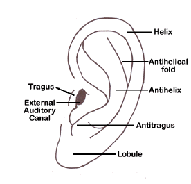
It is important to look at the ear structure and determine which components any malformation may involve. The image to the left shows the normal anatomy and structures of the external ear.
Ear position is also important, particularly if they are felt to be posteriorly rotated or low-set.
Please note: images that have a white symbol at the top right, such as the Preauricular Tags image below, indicates an image gallery that has multiple images - click on the image to open the gallery.
Preauricular Pits

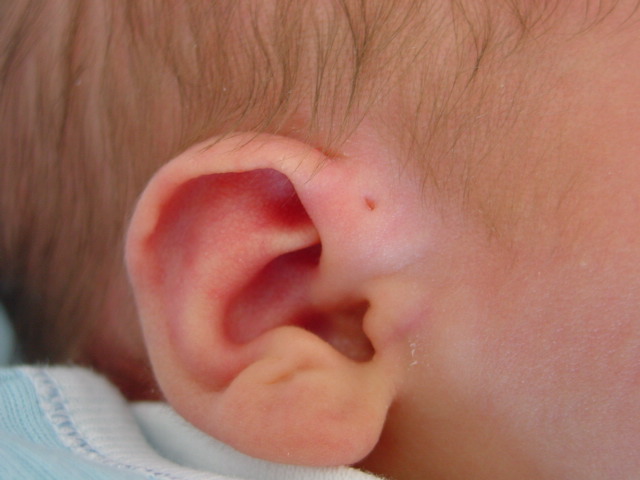
Preauricular pits are relatively common and are usually detected at birth. Some are complicated by infection as the child grows, and may need surgical attention at a later stage.
While usually benign (and often familial), they may be associated with other ear and face anomalies and may be present in branchio-otorenal dysplasia (with external ear anomalies, branchial fistulae, hearing impairment, and renal anomalies).
However, routine audiology testing is not indicated unless there are other risk factors for deafness.
Preauricular Tags
These are most often benign, isolated minor anomalies, occurring mostly unilaterally but occasionally bilaterally.
It is important however to examine the anatomic landmarks carefully. If the tags are associated with distortion of the pinna as noted in the photograph of the left ear then it should trigger suspicion of associated pathology such as the possibility of hemifacial microsomia.
Tags may also be seen as part of multiple dysmorphic features of infants with chromosomal anomalies. However, if isolated, no investigations are required and audiology referral is not necessary unless there are other risk factors, particularly a family history of hearing loss.
Pedunculated tags can be removed by tying them at the base should the parents wish but the parents need to be informed that tying the knot causes pain for a little while and will result in the tag changing colour, shrivelling up, and eventually falling of over the next week or so. Even if a tag is tied off, there may be a residual lump of soft tissue which may need subsequent cosmetic attention.
Microtia

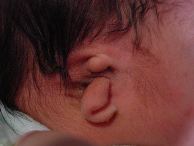
Microtia is a term used for abnormalities of ear development, ranging from subtle deviations of ear size, shape, and location of the pinna and ear canal, through to major malformations of the external ear with only small nubbins of cartilage and an absent auditory meatus. Anotia is complete absence of the pinna and ear canal.
Microtia-anotia is a rare condition, reported to occur in approximately 1.5:10,000 deliveries 1 (although it may be more common in some populations). About two-thirds of cases are isolated malformations, but it may occur in association with other malformations, and with specific syndromes in 10% of cases such as Treacher-Collins syndrome and Goldenhar (Hemifacial Microsomia) syndrome. Sporadic microtia is most likely to be unilateral; syndromic forms or those associated with other major malformations are more commonly bilateral.
Assessment in the neonatal period involves thorough examination for other abnormalities. It is prudent to check that the fetal anatomy scan was normal.
Although the parental concern will primarily be whether the ear will look cosmetically normal, the emphasis is initially on ensuring that the child has hearing sufficient for normal speech. Cosmetic repair will not be undertaken for several years, and is often done in combination with the otorhinolaryngology and plastic surgical services (there is a multidisciplinary microtia-anotia clinic at Starship Hospital).
Reference
Mastroiacovo P, Corchia C, Botto LD, Lanni R, Zampino G, Fusco D. Epidemiology and genetics of microtia-anotia: a registry based study on over one million births. J Med Genet 1995;32:453-7.
